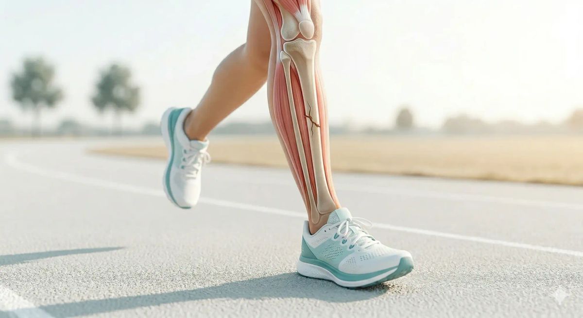
What is a bone stress injury?
What they are, how they are diagnosed, who is at risk, and what return to sport looks like, based on the published evidence.

What is a bone stress injury?
Bone is a living tissue. Under repetitive mechanical loading, it undergoes a continuous cycle of breakdown and rebuilding. When loading outpaces the bone's capacity to repair itself (through too much activity, too quickly, with insufficient recovery), microscopic damage accumulates. This is a bone stress injury (BSI).
The term "stress fracture" refers specifically to the late end of a spectrum. Modern clinical and research literature uses bone stress injury to cover the full continuum from early reaction to complete fracture.
The bone stress continuum
GRADE 1
Mild periosteal oedema on MRI. No symptoms at rest.
GRADE 2
Periosteal + early bone marrow oedema. Activity pain.
GRADE 3
Significant marrow oedema on T1 and T2. Pain at rest.
GRADE 4A
Unicortical fracture line. Considerable pain and disability.
GRADE 4B
Bicortical fracture line. May require offloading or surgery.
At the early end (grades 1, 2), the bone is reacting to load: swollen and stressed but not cracked. At the severe end (grades 4a, 4b), a distinct fracture line is present. This distinction matters because it drives management: simply, injuries caught promptly recover faster.
Where do they occur?
The lower limb accounts for the overwhelming majority of BSIs in runners. Common sites, in approximate order of frequency, include: tibia (posteromedial cortex), metatarsals (most commonly 2nd and 3rd), fibula, navicular, femoral shaft, femoral neck, calcaneus, and sacrum. Upper limb stress fractures are uncommon and tend to occur in throwing athletes or gymnasts.
- Femoral neck (tension side): Risk of complete fracture and avascular necrosis; may require surgical fixation.
- Anterior tibial cortex ("dreaded black line"): Poor healing potential; may require intramedullary nailing.
- Tarsal navicular: Central third is a watershed zone (poor blood supply); high non-union risk.
- Fifth metatarsal (Jones zone): Distinct from distal avulsion; higher surgical rate.
- Sacrum and pelvis: Often missed; highly relevant in athletes with suspected RED-S.
Presentation
The typical history is activity-related pain that is well-localised to a bony site, developing gradually over two to three weeks following a change in training load. Initially, the pain occurs only during activity; in more severe injuries, it persists at rest or disturbs sleep. Point tenderness directly over bone (not over muscle or tendon) is the key examination finding. The hop test, which involves single-leg hopping on the affected limb, reproduces pain in tibial and lower limb injuries.
Imaging
| Modality | Role | Limitation |
|---|---|---|
| Plain X-ray | First-line; low cost; may show periosteal reaction or fracture line at later stages. | Normal in up to 70% of early stress fractures. |
|
MRI
Gold Standard |
Detects all grades; visualises periosteal and marrow oedema before fracture develops. | Requires MSK specialist interpretation. |
| CT | Useful for precise fracture line characterisation (e.g. navicular, anterior tibia). | Ionising radiation; inferior to MRI for early marrow changes. |
| Bone Scintigraphy | Historically used; highly sensitive after 72 hours. | Low specificity; largely superseded by MRI. |
Plain X-ray remains the appropriate first investigation given availability. If X-ray is normal but clinical suspicion is high, MRI is the next investigation of choice. For high-risk sites like the femoral neck or navicular, do not wait for X-ray confirmation before offloading the limb.
Training load: the single-session spike
A 2025 cohort study by Frandsen et al. in the BJSM followed 5,205 runners and challenged the "10% weekly rule." They found that neither the week-to-week ratio nor the cumulative workload (ACWR) showed a significant association with injury risk. The single-session spike did.
Runners who completed a session more than 10% longer than their longest run in the preceding 30 days had a 64% higher rate of overuse injury (hazard rate ratio 1.64, 95% CI 1.31 to 2.05).
While these findings are powerful, the study was observational. This means it tracked the natural patterns of runners rather than controlling every variable in a lab. Factors like your personal injury history, nutrition, and running style still play an important role. However, the data strongly suggests that avoiding these "distance spikes" is one of the most effective ways to protect your bones.
Risk factors
Risk factors for BSI are broadly categorised as intrinsic (factors specific to the individual’s biology or biomechanics) or extrinsic (environmental or training-related factors).
Intrinsic: Biomechanical
- High vertical loading rate
- Reduced cadence / increased stride length
- Low bone mineral density
- Leg length discrepancy
Intrinsic: Nutritional & Hormonal
- Low energy availability (RED-S)
- Amenorrhoea / Menstrual dysfunction
- Vitamin D or Calcium deficiency
- Low BMI (< 21 kg/m²)
Extrinsic: Training
- Single-session distance spike (> 10%)
- Abrupt transition to harder surfaces
- Insufficient recovery days
- Rapid increase in overall volume
Extrinsic: Equipment
- Carbon-plated "Super Shoes" (potential navicular strain)
- Worn or inappropriate footwear
- Cambered running surfaces
Relative Energy Deficiency in Sport (RED-S)
Athletes with low energy availability face a substantially higher risk of BSI. Those at moderate RED-S risk are approximately twice as likely to sustain a BSI; those at high risk, approximately four times as likely. Amenorrhoeic athletes carry a two to four fold higher risk compared with those with regular menstrual function. RED-S affects male athletes as well as female, though the evidence base in males is less established.
Management
Load modification is the cornerstone of management. This does not necessarily mean complete rest. It means removing the offending mechanical stimulus while maintaining fitness through low-impact alternatives (pool running, cycling, swimming) as pain permits.
For high-risk sites (femoral neck, navicular, anterior tibia), protected weight-bearing with crutches is indicated until the diagnosis is confirmed and a specialist plan is in place. Surgical review should not be delayed.
For low-risk sites (posteromedial tibia, fibula, metatarsals 2–4), management is largely conservative: relative rest, progressive reloading guided by pain response, and correction of contributing factors like vitamin D status or footwear.
Successful long-term recovery involves more than just rest; it requires identifying and fixing the underlying cause. This includes correcting training mistakes, checking footwear, and ensuring proper intake of Vitamin D and Calcium. For athletes with repeat injuries, low body weight, or irregular periods, a specialist may recommend a DEXA bone density scan to assess overall bone health and screen for energy deficiency (RED-S).
Return to sport timelines: Low-risk sites
Grade 1–2: Stress Reaction
2–4 weeks
Activity modification; transition to pain-free running when periosteal tenderness resolves and "Hop Test" is negative.
Grade 3: Moderate BSI
4–8 weeks
Relative rest from running; focus on cross-training; gradual return programme.
Grade 4A: Unicortical Fracture Line
8–12 weeks
Protected weight-bearing may be required; specialist-guided return.
Grade 4B: Bicortical Fracture / High-Risk Site
12+ weeks: variable
Surgical consultation likely; femoral neck fractures may require internal fixation.
Return to running should follow a structured programme, beginning with walk–run intervals and progressing based on pain response. A graduated return typically spans 4–6 weeks from initial reintroduction to full training. Imaging-based clearance before return is not routinely required for low-risk sites but should be considered for high-risk anatomical locations.
When to seek assessment
Seek specialist assessment if you have: localised bony tenderness following a change in training load; groin or hip pain (to exclude high-risk femoral neck injury); midfoot pain that is tender over the navicular; recurrent stress injuries; or any suspected high-risk site injury. Early diagnosis significantly shortens recovery time.
EVIDENCE BASE
Frandsen JSB, et al. Identifying high-risk running sessions. Br J Sports Med. 2025;59(17):1203–1210.
Nattiv A et al. MRI grading of bone stress injuries. Am J Sports Med. 2013.
Mountjoy M et al. IOC consensus statement on RED-S: 2018 update. Br J Sports Med.